Figure 5.

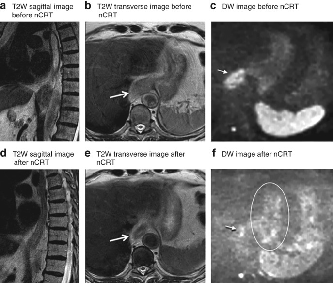
MRI of a patient with locally advanced oesophageal cancer located at the gastro‐oesophageal junction Images from an 80‐year‐old man with a cT3 N0 squamous cell carcinoma located at the gastro‐oesophageal junction. Histopathology after oesophagectomy showed residual tumour (tumour regression grade 2, ypT1a N0). a–c T2‐weighted (T2W) sagittal (a) and transverse (b) images before neoadjuvant chemoradiotherapy (nCRT) show a thick hyperintense wall, accompanied by a hyperintense signal on diffusion‐weighted (DW) imaging (c). d–f After nCRT, the T2W images (d,e) show shrinkage of the wall with a mixed hyperintense and hypointense signal, which was assigned a confidence level score of 3 by all readers. The DW image (f) shows spots of hyperintense signal in the primary tumour area (arrow), which is suspicious for residual tumour and was therefore assigned a confidence level score of 4 by all readers. The area within the circle indicates normal stomach wall, which also shows small hyperintense areas on DW imaging. Arrows indicate tumour location.
