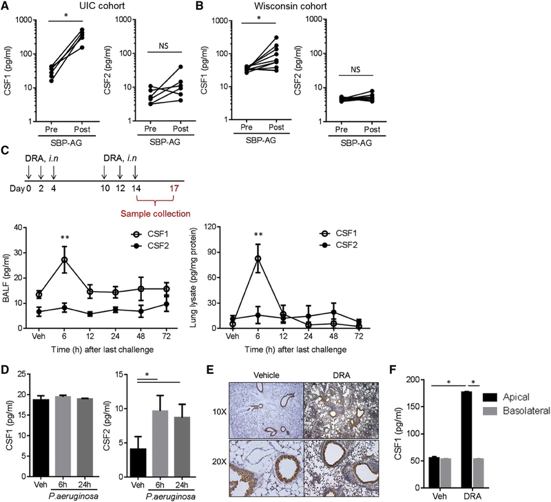
Figure 1. Airway epithelial cells secrete CSF1 into the airspace in response to aeroallergen challenge.
(A) CSF1 and CSF2 in BAL fluids obtained from patients with mild asthma enrolled in the UIC SBP-AG cohort (n=6). (B) CSF1 and CSF2 in the Wisconsin SBP-AG cohort (n=10). (C) Schematic of the DRA-induced mouse asthma model. DRA allergens were given intranasally (i.n.). CSF1 and CSF2 in BAL fluids (left panel) and lung lysates (right panel) in the DRA model (n=5). (D) Concentrations of BAL CSF1 & CSF2 in P. aeruginosa lung infection model (n=3). (E) Anti-CSF1 immunohistochemical (IHC) staining with lung tissues from the mice with DRA or vehicle only. (F) CSF1 in the transwell media of primary bronchial epithelial cells (BEAS-2B) stimulated with DRA allergens (each 1 μg/ml) for 24h. The media from apical and basal chambers were independently collected. Data represent at least two (C-E) or three (F) independent experiments * p<.05, **p<0.01. Please also see figure S1A–D.

