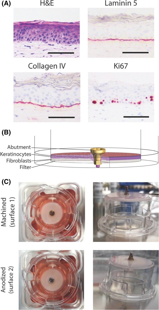
Figure 1.

The reconstructed human gingiva (RHG) implantation model. A, Immunohistochemical analysis of paraffin embedded tissue sections is shown. Magnification bar = 100 μm. B, A scheme showing the experimental design. C, A macroscopic view of the implants placed into the RHG. The RHG is shown at the time of harvesting, which was after 10 days of culture at the air‐liquid interface. The transwell diameter = 2.5 cm. Representative results obtained from 6 images and 3 independent experiments are shown; see Materials and Methods, section Data Analysis for further information
