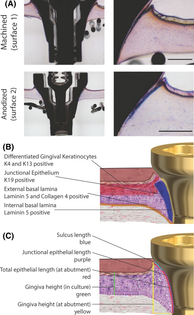
Figure 2.

Histomorphometric analysis shows reconstructed human gingiva (RHG) attached to machined (surface 1) and anodized (surface 2) surfaces. A, Tissue sections 80‐100 μm thick were surface‐polished and surface‐stained with McNeal's Tetrachrome, basic Fuchsine, and Toluidine blue. Representative results obtained from 12 images and 3 independent experiments are shown; see Materials and Methods, section Data Analysis for further information. Scale bar = 500 μm. B, A schematic representation of the RHG implantation model, which indicates tissues of interest and their protein expression. C, A schematic representation of the RHG implantation model with a visualization of parameters that were measured histomorphometrically
