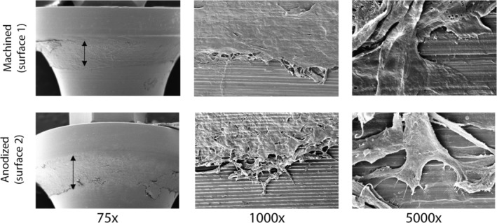
Figure 3.

Scanning electron microscopy showing epithelial cell attachment to the abutment surfaces. Left: double headed: The arrow indicates the width of the attached epithelium. Middle: An example of the migrating epithelial front. Right: An example of keratinocyte attachment to the abutment surface. Numbers indicate the fold magnification. Representative results obtained from 3 independent experiments are shown; see Materials and Methods, section Data Analysis for further information
