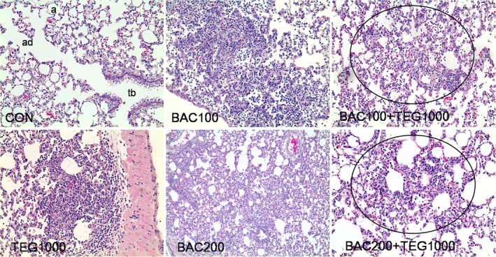
Figure 8.

Histopathology of lung tissues from rats intratracheally exposed to BAC and TEG. Male rats were intratracheally instilled with BAC (100 or 200 μg/kg) and TEG (1000 μg/kg), either individually or together. Lung tissues were isolated and stained with H&E after 1 day exposure. Multifocal bronchiolar/alveolar acute inflammation in the circle. tb, terminal bronchioles; ad, alveolar ducts; a, alveoli. H&E. Magnification, 100× for BAC200 and 200× for others [Color figure can be viewed at wileyonlinelibrary.com]
