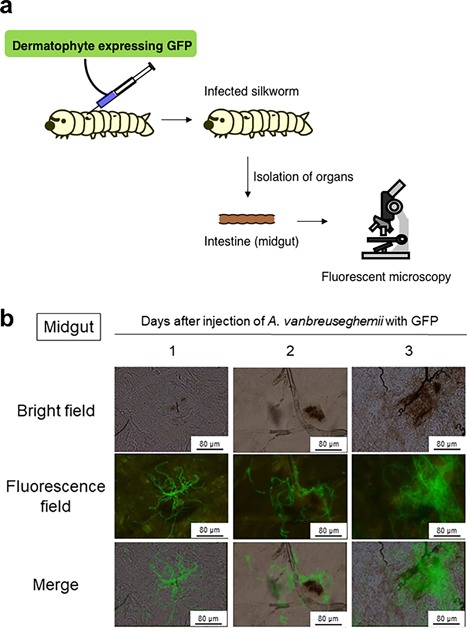
Figure 3.

Visualization of infection in organs of silkworm using a dermatophyte expressing eGFP. (a) Method for evaluation of hyphal growth of dermatophytes in organs of silkworm by fluorescent imaging. (b) Fluorescence microscope images of midgut of silkworm infected with A. vanbreuseghemii expressing eGFP . Figure 3b was reproduced from Ishii et al., (8) with permission.
