
Figure 1.
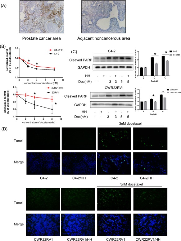
CD4+ T‐cell infiltration in PCa patients with Doc treatment and altered PCa chemotherapy sensitivity. A, Clinical specimen IHC staining from Doc treatment PCa patients showed that more CD4+ T cells infiltrated the prostate cancer area than in the adjacent noncancerous area. B, PCa cells were cocultured with HH cells for 2 days in 24‐well plates under 0.4 μm transwell membranes; a total of 1 × 10⁵ PCa, C4‐2/CWR22Rv1, were plated in lower chambers of transwells and 1 × 10⁵ CD4+ T cells, HH were plated on the upper insert chambers. Subsequently, cells were treated with different doses of Doc for 48 hours and tested with CCK8. Consider absorbance of 0 nM as control, all absorbance of other doses was compared to control. After coculture with HH cells, C4‐2 cells become more resistant to Doc treatment. Data are presented as mean ± SD, n = 3. *P < 0.05 vs control. C, Western blot analysis: for Doc treatment, PCa cells cocultured with HH cells showed less expression of cleaved PARP compared to PCa cells without coculture HH cells. Data are also presented as mean ± SD, n = 3. *P < 0.05 vs control. D, Tunel assay analysis of cell apoptosis. For Doc treatment, PCa cells cocultured with HH cells showed less cell apoptosis compared to PCa cells without coculture HH cells. CCK8, cell counting kit‐8; Doc, Docetaxel; GAPDH, glyceraldehyde 3‐phosphate dehydrogenase; IHC, immunohistochemistry; PARP, poly ADP ribose polymerase; PCa, prostate cancer; Tunel, terminal deoxynucleotidyl transferase dUTP nick‐end labeling [Color figure can be viewed at wileyonlinelibrary.com]