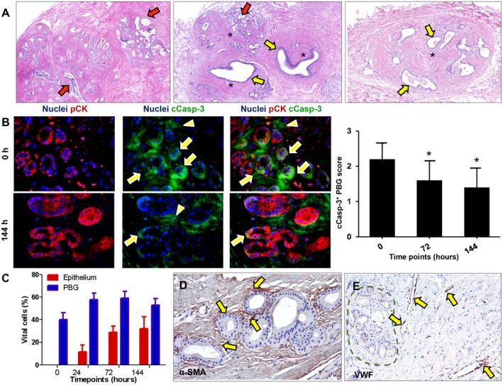
Figure 2.

Bile ducts that were subjected to severe ischemia and subsequent reoxygenation showed cell death and PBG dilatation. (A) Hematoxylin and eosin staining showing necrotic PBG leaving cell debris in the empty acini (red arrows). In addition, PBG dilatation occurred in specific glands (yellow arrows). In some sections, both cell death and dilatation were present in proximate PBG collections (central image). Some PBG collections were captured in a concentric formation of stromal cells (asterisks). (B) Immunofluorescence for pCK and cCasp‐3 indicated significantly less apoptosis in PBG cells after 72 hours and 144 hours of incubation compared with baseline (*P < 0.05). Cleaved caspase‐3 was expressed by the PBG cells (arrows) as well as around the PBG cells in the stroma (arrowheads). Nuclei are displayed in blue. (C) Quantification of the percentage of vital cells in PBG and epithelium showing that approximately 50% of the PBG cells appeared vital during all time points, with no significant differences over time. In contrast, no luminal epithelial cells were present at baseline, yet a growth of epithelium up to 32% apparent vital cells was evident up to 144 hours of incubation. (D) Immunohistochemistry for α‐SMA illustrates presence of myofibroblasts around PBG and provides evidence for an anatomical organization of stromal cells (arrows). (E) Immunohistochemistry for VWF in endothelial cells (arrows) indicating vessels throughout the stroma in proximity of PBG (dotted line). Dilated vessels with loss of endothelial cells can be observed. (A‐C) Original magnification ×10. (D,E) Original magnification ×20. Abbreviations: α‐SMA, alpha‐smooth muscle actin; cCasp‐3, cleaved caspase‐3; h, hours; pCK, pan cytokeratin; VWF, von Willebrand factor.
