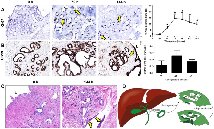
Figure 5.

PBG proliferation and regeneration after ischemia‐induced epithelial cell loss. (A) Immunohistochemistry for Ki‐67 showed mitotic activity after 24 hours of incubation. PBG proliferation index increased significantly after 24 hours, peaked at 72 hours, and remained constant until 144 hours of incubation, compared with baseline (*P < 0.05). (B) Immunohistochemistry for biliary epithelial marker CK19 showed that PBG and biliary epithelium expressed CK19 during all time points. The pattern of the relative number of CK19 expressing cells resembled that of proliferation. (C) Hematoxylin and eosin staining displaying complete loss of epithelial layer at baseline and regeneration of an epithelial monolayer after 144 hours of incubation. This monolayer appeared at the surface of an open place within the stroma (yellow arrows). (D) Schematic overview and summary of the observed biliary damage and cellular reaction after ischemia and subsequent reoxygenation. After ischemia and reoxygenation, the biliary epithelium was generally detached and PBG were necrotic. Directly after reoxygenation, PBG showed cell debris in the acini with a few residual PBG cells. After 6 days of oxygenation, proliferation, refilled PBG, and regeneration of the epithelium were evident. (A) Original magnification ×10. (B,C) Original magnification ×20. Abbreviations: h, hours, L, lumen.
