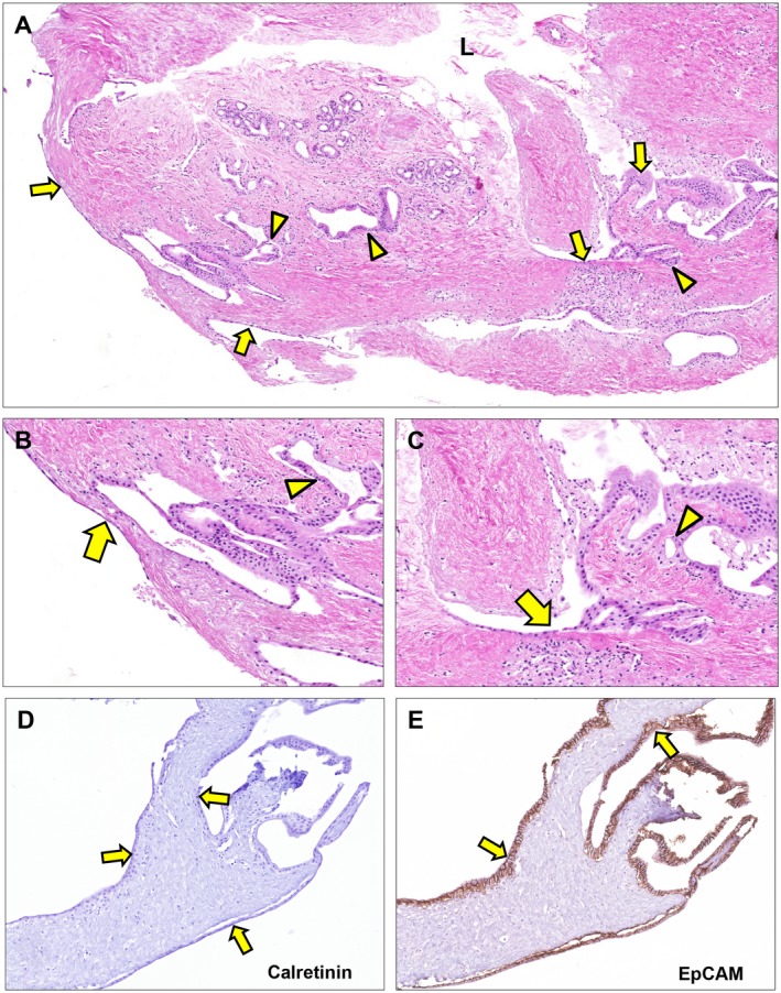
Figure 6.

Epithelial regeneration after 72 hours of ex vivo culture. (A‐C) Hematoxylin and eosin staining. Epithelial lining appeared at the luminal surface as well as at the basolateral surface (arrows). These epithelial patches are in proximity of PBG (arrowheads) suggesting newly formed epithelium driven from PBG to restore the uncovered surface at several sides throughout the tissue slice. (D) Epithelial lining at basolateral and luminal side of the bile duct slice appeared negative for calretinin, which is specific for mesothelium (arrows). (E) All cells lining the basolateral and luminal side of the bile duct slice expressed EpCAM, confirming that the (re)generated monolayers consisted of epithelial cells (arrows). (A) Original magnification ×5. (B,C) Original magnification ×20. (D,E) Original magnification ×15. Abbreviation: L, lumen.
