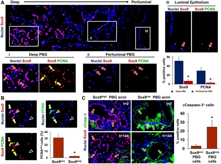
Figure 8.

Immunophenotype of deep and periluminal PBG. (A) Immunofluorescence for Sox9, which identifies endoderm‐derived progenitor cells, showed that PBG located in deeper position (i) with respect to the luminal epithelium were characterized by higher expression of Sox9 compared with PBG acini located in a periluminal position (ii) and in continuity with luminal epithelium (iii). Sox9+ cells (red arrows) coexpressed the proliferation marker PCNA, merely in the deeper‐located PBG (i: yellow arrows). Periluminal PBG showed less Sox9 expression and almost no PCNA expression (ii: red arrows). The luminal epithelium (iii) showed rare Sox9+ cells (red arrow) and negligible levels of PCNA. The corresponding graph shows that both Sox9 and PCNA expression were significantly higher in deeper PBG, compared with periluminal PBG. (B) Specifically, PBG acini with Sox9‐ cells expressed no PCNA (black arrowheads) and acini with abundant Sox9 expression stained positive for PCNA (yellow arrows). Red arrowheads point toward Sox9+ cells that were PCNA negative. Almost all PCNA+ cells were Sox9+ as displayed in the graph, confirming that the proliferating cell population consisted of mainly progenitor cells. (C) PBG harboring Sox9+ cells (red arrows) were less positive for the apoptosis marker cCasp‐3. PBG harboring mainly Sox9‐ cells and a few Sox9+ cells (yellow arrow) showed marked expression of cleaved caspase‐3 (green arrows). This was evident during all time points. Quantification of Sox9/cCasp‐3 coexpression revealed significantly higher expression of cCasp‐3 in Sox9‐ PBG cells, compared with Sox9+ PBG cells. (A) Original magnification ×10. Area in the boxes is magnified at ×20. (B,C) Original magnification ×20. Abbreviation: cCasp‐3, cleaved caspase‐3.
