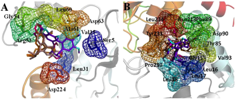
Figure 2.
The active pockets of protein UGT1A1 (A) and UGT1A3 (B) for binding with schisandrin A, schisandrin, and schisandrin C. The C atoms of schisandrin A are colored with oranges, and N atoms colored with magenta. The C atoms of schisandrin are colored with purple, and N atoms colored with cyans. The C and N atoms of schisandrin C show violet and hot pink, respectively. All the three inhibitors are shown in stick, and the residues in the active pockets of proteins are shown in stick and mesh. This figure is available in color online at wileyonlinelibrary.com/journal/ptr.

