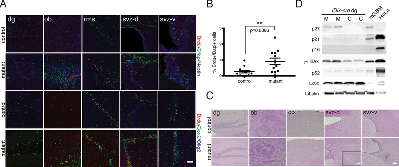
Figure 5. iDlx-cre Mutants exhibit Hyperplastic Lesions and Molecular Phenotypes.
A. Immunoflourescence staining of iDlx-cre mutant vs. control with BrdU (after a short term pulse) and lineage markers (svz-d=dorsal SVZ region, svz-v=ventral SVZ region). B. Quantification of % BrdU-positive cells in iDlx-cre mutants (n=12) vs. controls (n=12). p=0.0086. **p<0.01 using two-tailed unpaired Student’s t-test. Data is presented as mean +/− SEM. C. H&E staining of aged iDlx-cre mutant vs. control mouse brains reveal regions of hypercellularity (>28 weeks post-induction). Inset in mutant svz-d showing magnified lateral ventricle. D. Western blot analysis of iDlx-cre mutants and controls using DNA damage (gH2Ax), senescence (p16, p21, p27) and autophagy (Lc3b, p62) markers. Mouse GBM (mGBM) and HeLa cell lysates were used as controls. All scale bars=100 μm. In A and C-D, experiments were independently repeated with similar results at least n=3 times using at least n=3 different mouse tissue samples for each group

