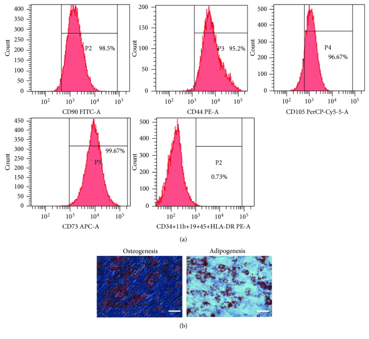
Figure 1.
Characterization of hAMSCs. (a) FACS analysis of cell markers of hAMSCs. hAMSCs were >90% positive for CD105, CD73, CD44, and CD90 and negative for CD34, CD11b, CD19, CD45, and HLA-DR. (b) Differentiation of hAMSCs into osteocytes and adipocytes. Cells cultured under osteogenic or adipogenic culture conditions were stained for calcium deposits with Alizarin red staining or lipid droplets with Oil Red O staining. Scale bar = 50 μm. hAMSCs: human amniotic mesenchymal stem cells.

