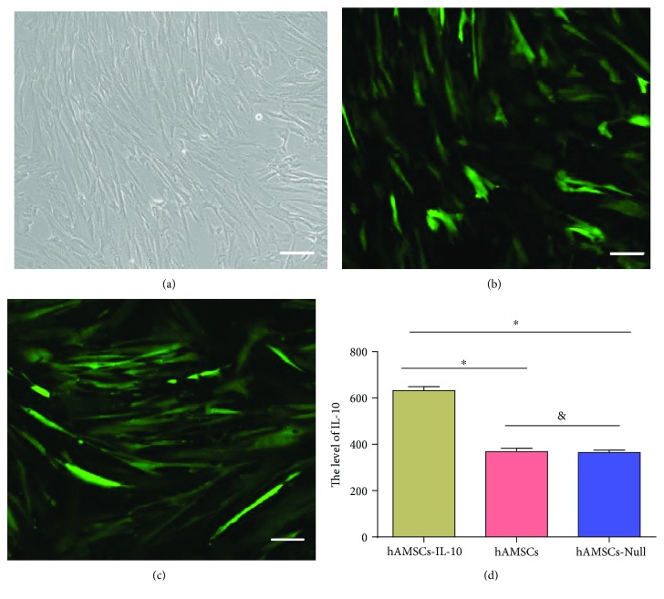
Figure 2.
Morphology, transfection efficiency, and IL-10 expression of hAMSCs. (a) Phase-contrast micrograph of hAMSCs showing spindle-shaped morphology. (b) Fluorescence micrographs of hAMSCs after LV-IL-10 infection for 48 h. (c) Fluorescence micrographs of hAMSCs after LV-Null infection for 48 h. (d) IL-10 expression of hAMSCs; the concentration of IL-10 from hAMSCs-IL-10 increased compared to that of hAMSCs-Null and hAMSCs. ∗P < 0.05 and &P > 0.05. LV-IL-10: replication-defective lentivirus expressing IL-10; LV-Null: replication-defective lentivirus not carrying any exogenous genes. Scale bar = 500 μm.

