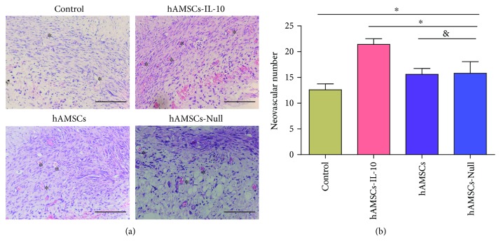
Figure 6.
Effects of hAMSCs on wound neovascularization on day 7 after cell transplantation. (a) H&E staining showed more new blood vessels in the hAMSCs-IL-10, hAMSCs, and hAMSCs-Null groups than in the control group. The hAMSCs-IL-10 group had the most new blood vessels, and the hAMSCs-Null and hAMSC groups were not significantly different. ∗New blood vessel. Scale bar = 500 μm. (b) Quantification of new blood vessels in the dermis by groups on day 7. The number of new blood vessels in the dermis increased after treatment with hAMSCs-IL-10 compared with hAMSCs, hAMSCs-Null, or control on day 7 and increased with hAMSCs and hAMSCs-Null compared with the control. No significant differences between the hAMSCs and hAMSCs-Null groups were seen in vascularization on day 7. ∗P < 0.05 and &P > 0.05.

