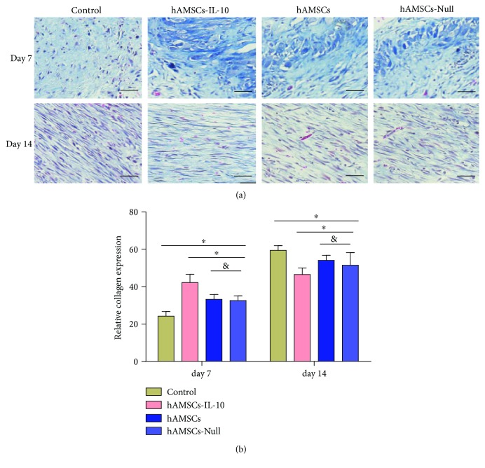
Figure 8.
Collagen accumulation in wounds on days 7 and 14. (a) Masson staining of collagen deposition in the dermis showed increases in the hAMSCs-IL-10, hAMSCs, and hAMSCs-Null groups on day 7 compared with the control group; the hAMSCs-IL-10 group had the most collagen, and collagen was arranged regularly; the hAMSCs-Null and hAMSC groups were not significantly different. On day 14, the control group had the highest collagen deposition, and collagen was arranged irregularly. The hAMSCs-IL-10 group had the lowest collagen deposition. Scale bar = 50 μm. (b) Quantification of collagen synthesis in wound skin by groups at indicated time points.∗P < 0.05 and &P > 0.05.

