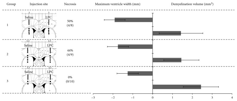
Figure 2.
Optimization of the LPC injection protocol. LPC concentration (1% in saline) and infusion rate (0.1 μl/min) were kept constant between the three groups. The injection sites are shown in the corresponding Paxinos coronal diagram, with the following coordinates: group 1, AP −0.3 mm, ML ±3.0 mm, DV −3.5/−4.0/−4.5/−5.0 mm; group 2, AP −0.3 mm, ML ±3.3 mm, DV −3.0/−3.7/−4.3/−5.0 mm; group 3, AP −0.3 mm, ML ±3.3 mm, DV −2.8/−3.5 mm. Each site was infused with 2.5 μl of LPC, from depth to superficial. The rate of animals exhibiting focal edematous hypersignals on MRI (as in Figure 1(b)) is given as a percentage (and number of animals out of total the group number). The graph shows the maximum ventricle width (measured along the mediolateral plane as in Figure 1(c), and arbitrarily expressed as a negative value, in mm) and the total volume of corpus callosum exhibiting a normalization of the natively hypointense contrast (measured as in Figure 1(a), in mm3).

