Figure 10.

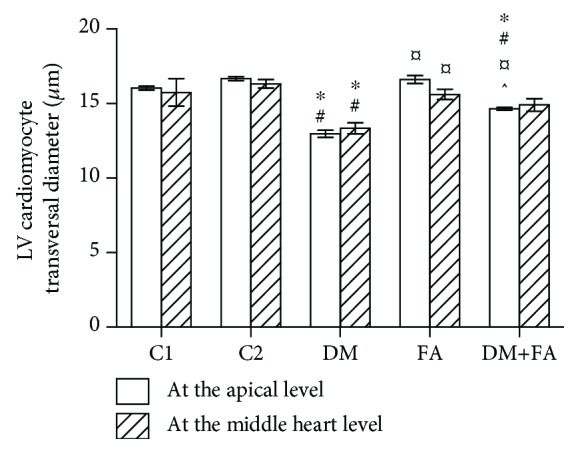
Left ventricular (LV) cardiomyocyte transversal diameter measured at the apical and middle heart levels. ∗P < 0.05 versus the C1 group, #P < 0.05 versus the C2 group, ¤P < 0.05 versus the DM group, and ^P < 0.05 versus the FA group.

Left ventricular (LV) cardiomyocyte transversal diameter measured at the apical and middle heart levels. ∗P < 0.05 versus the C1 group, #P < 0.05 versus the C2 group, ¤P < 0.05 versus the DM group, and ^P < 0.05 versus the FA group.