Figure 1.
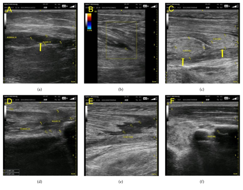
Grade I muscle rupture, but focal fibre rupture with less than 5% of the muscle involved; ultrasonography shows a hypoechoic mass within the muscle fibres (yellow arrow) (a, b). Grade II partial muscle rupture; the muscle belly forms a real mass, with focal fibre rupture with more than 5% but less than 30% of the muscles involved. Ultrasonography shows a hypoechoic mass within the muscle fibres (yellow arrow) (c, d). Grade III lesion represents focal fibre rupture with more than 30% of muscle rupture with retraction, and it also shows the bell-clapper sign, with torn muscle fragments floating in the serohematic fluid (yellow arrow) (e). Ultrasonography shows a limb fracture, fracture of the radius (yellow arrow point) (f).

