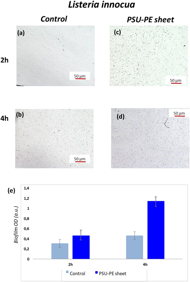
Figure 5.

(a,b) Optical microscopy images of L. innocua bacterial adhesion at two different time points on control and (c,d) on the polysulfone pyro-electrified (PSU-PE) sheet; (e) histogram of bacteria biofilm OD (optical density) on the control and the PSU-PE sheet at two different time points.
