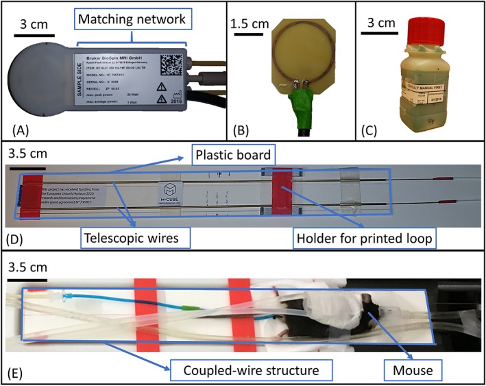
Figure 2.

Coupled‐wire inspired coil for 7 T small animal imaging: (A) picture of the commercial surface coil (Bruker, model 1P T957 8 V); (B) picture of the printed loop of 3 cm diameter on circuit board (FR‐4, 0.5 mm thickness) and fed through a coaxial cable; (C) Bruker 19F/1H phantom of 3.5 x 3.5 x 9 cm3 used during on‐bench measurements and MRI scans, concentration of 19F of 100 mmol/L; (D) picture of the coupled‐wire structure with two telescopic wires separated by 3 cm and placed on a plastic board; (E) picture of the in vivo experimental setup
