
Figure 2. ECFC adhesion increases on biomolecule-modified PVA.
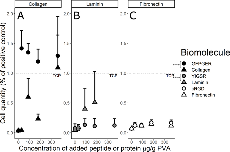
ECFC quantification after 48hrs on PVA films for the (A) collagen, (B) laminin, and (C) fibronectin families demonstrated that cell attachment increased on the biomolecule-modified PVA compared to the plain PVA. Data were analyzed with a 2-way ANOVA to compare each peptide to its corresponding protein. Factors were biomolecule and concentration. GFPGER showed a significant increase (as indicated by * in the figure) in ECFC binding compared to collagen modification (ANOVA F1,56=27.04, p=2.91×10⁻⁶), and laminin showed a significant increase from YIGSR (ANOVA F1,75=12.84, p=0.0006). Fibronectin and cRGD were not significantly different (ANOVA F1,88=3.07, p=0.0831). As a factor, increasing concentration significantly increased cell attachment in the collagen (ANOVA F1,56=22.29, p=1.61×10⁻⁵) and fibronectin (F1,88=13.04, p=0.0005) families, but not in the laminin family (F1,75=1.51, p=0.224).