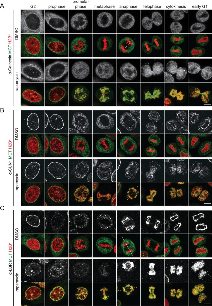
FIGURE 2:
ER and NE membrane proteins are distributed throughout the mitotic ER in the presence of MCT-induced chromatin–membrane contacts. MCT/H2B* cells were treated as outlined in Figure 1B, immunostained using anti-calnexin (A), anti-SUN1 (B), and anti-LBR (C) antibodies, and analyzed by confocal microscopy. Chromatin configuration was used to assign cells to the indicated cell cycle stages. Dashed lines represent spindle axes. Bars, 10 μm.

