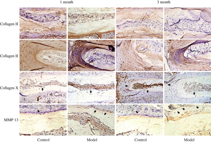
Figure 4.

Immunohistochemical assessments of the protein level of type II collagen, type X collagen, and MMP13. Immunohistochemical study showed that the protein level of type II collagen was decreased slightly after modeling in both 1 month and 3 month groups compared with the control group. The collagen X positive staining reached to the outer annulus fibrosus and the nucleus pulposus, and that of the 3‐month group was stronger than for the 1‐month group. The positive staining of MMP‐13 of the 1‐month and 3‐month groups reached to the edge of the annulus fibrosus and nucleus pulposus, and was stronger for the 3‐month group than for the 1‐month group.
