Figure 2.

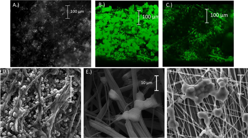
A.) Micrograph showing an insert with PC12 cells immobilized onto the fibers (cells stained with acridine orange); B-C.) Confocal images taken of a section of the collage-coated PS inserts. B.) Micrograph showing clumped cells on top with cells in fibers below. C.) Same view of the cells with the clumped cells on top removed. D.-F) Scanning electron microscopy images showing PC12 fixed onto PS fibers. D.) Well split groups of cells permeate the fibers throughout. E.) Clumps of cells branching out to hold the substrate. F.) Cells bunching together on the surface of a thin sheet of fibers.
