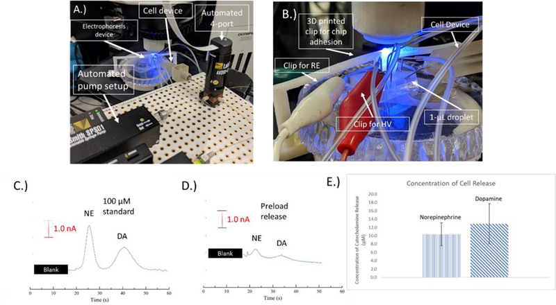
Figure 5.

A.) Picture of the electrophoresis setup. The cell device can be seen connected with a capillary to the automated syringe and valve system. The device is connected to the bilayer valving chip. B.) Picture showing the integration of the cell device with the electrophoresis chip. The steel pin can be seen integrated with the 1-µL droplet. C-E.) Electropherograms of C.) 100 µM norepinephrine and dopamine D.) Detected release of norepinephrine and dopamine from pre-loaded PC12 cells E.) The release of norepinephrine and dopamine from cells pre-loaded with 1 mM of both for 1 hr prior to the experiments. E.) A bar graph showing a comparison of the norepinephrine and dopamine detected from the pre-loaded PC12 cells (n=4).
