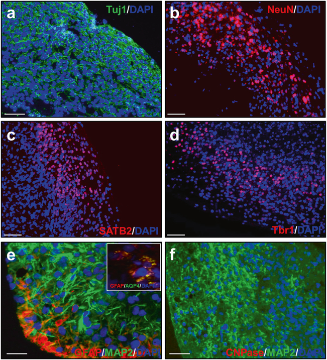
Fig. 2.
Cell types in COs at 110 days in culture. COs prepared from HC iPSCs showed that most of the cells express the neuronal markers Tuj1 (a) and NeuN (b). Additionally, the cells express the cortical markers SATB2 (c) and Tbr1 (d). e Some cells were positive for the astrocyte markers GFAP and aquaporin 4 (AQP4, inset), but no cells were positive for the oligodendrocyte marker CNPAse (f). DAPI in blue. Scale bar 50 μm in all panels

