Fig. 5.
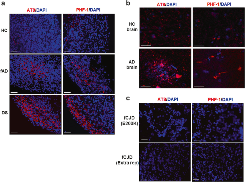
Accumulation of hyper-phosphorylated tau in COs. a COs prepared from iPSCs derived from healthy control (HC, top), familiar Alzheimer’s disease (fAD, center), and Down syndrome (DS, bottom) patients were cultivated for 110 days and processed for histological analysis as described in the Methods. Representative images of COs stained with AT8 (left) and PHF-1 (right), two antibodies that recognize p-tau protein. b Staining of p-tau with the same antibodies was done in slides coming from the brain of HC (top) and AD (bottom) patients. c To evaluate the specificity of p-tau accumulation, COs prepared from iPSCs derived from patients affected by two different forms of familial Creutzfeldt-Jakob disease (fCJD) were stained with AT8 (left) and PHF-1 (right). DAPI in blue. Scale bars: 25 μm

