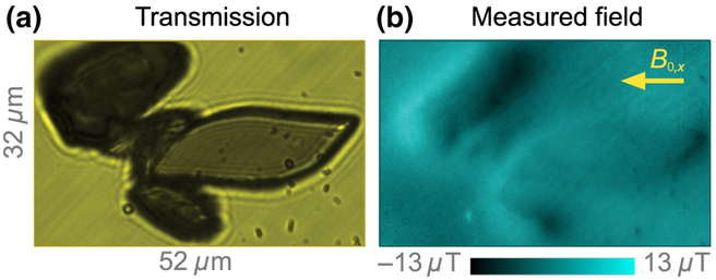
FIG. 11.
Magnetic imaging of hemin crystals. (a) Bright field transmission image of hemin crystals. (b) Corresponding diamond-magnetic-microscopy image (B0 = 186 mT). The inplane component of the applied field is labeled with an arrow. An out-of-plane component of similar magnitude is also present. Smaller particles are synthetic hemozoin crystals that are dispersed along with hemin crystals.

