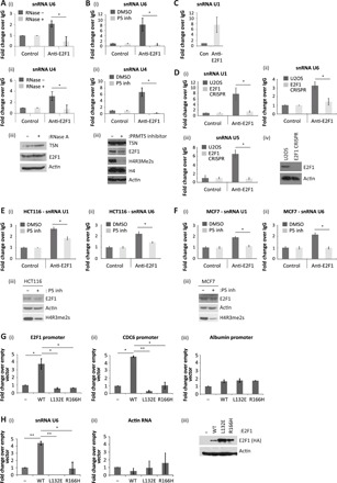
Fig. 3. E2F1 interacts with components of the splicing machinery.

(A) U2OS cells were lysed in RIP lysis buffer, containing ribonuclease A (RNase A; 20 μg/ml) where indicated. Cell extracts were immunoprecipitated with E2F1 antibody, and coimmunoprecipitated RNA was reverse-transcribed before quantitative polymerase chain reaction (qPCR) analysis with primers against U6 (i) and U4 (ii) snRNAs as indicated. Input protein levels were determined by immunoblot (iii). n = 2. (B) U2OS cells were treated with 5 μM PRMT5 inhibitor (P5 inh), as indicated, before performing an anti-E2F1 RIP. Coimmunoprecipitated U6 (i) and U4 (ii) snRNAs were identified with specific primers by quantitative reverse transcription PCR (qRT-PCR). Input protein levels were determined by immunoblot (iii). n = 3. (C) An anti-E2F1 RIP was performed on U2OS cells, and coimmunoprecipitated U1 snRNA was detected by qRT-PCR. n = 2. (D) An anti-E2F1 RIP was performed on extracts prepared from U2OS or U2OS E2F1 CRISPR cell lines as indicated. Immunoprecipitated RNA was analyzed by qRT-PCR using primers specific to U1 (i), U6 (ii), or U5 (iii) snRNAs. Input protein levels are also displayed (iv). n = 2. (E) HCT116 cells were treated with 5 μM PRMT5 inhibitor, where indicated, before performing an anti-E2F1 RIP. Coimmunoprecipitated U1 (i) and U6 (ii) snRNA were detected by qRT-PCR. Input protein levels are also displayed (iii). n = 2. (F) As described above, although the experiment was performed in MCF7 cells. (G) U2OS cells were transfected with 1 μg of plasmid encoding WT E2F1, DNA binding domain mutant constructs (L132E and R166H) or empty vector (−) as indicated. Forty-eight hours later, cell extracts were used for ChIP analysis with the anti–hemagglutinin (HA) antibody. Immunoprecipitated chromatin was analyzed by qPCR using primers targeting the indicated promoters, where albumin served as the non-E2F target gene control (i to iii). Input protein levels are shown in (H). n = 2. See also fig. S4B. (H) U2OS cells were transfected as above. Forty-eight hours later, cell extracts were used for RIP analysis with anti-HA antibody. Immunoprecipitated RNA was analyzed by qRT-PCR using primers specific to U6 snRNA (i) or actin RNA (ii). Input protein levels were determined by immunoblot (iii). n = 3.
