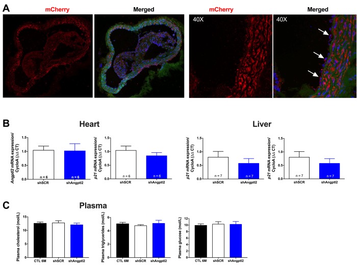
Figure 3.
Distribution of the AAV1-mCherry in the aortic wall and specificity of the AAV1-shAngptl2. (A) Immunofluorescence of AAV1-mCherry in frozen aortic sections of ATX mice at 6 months of age, 3 months post-infection: mCherry signal distributed throughout the vascular wall is shown in red and basal lamina in green; nuclei are shown in blue. At a higher magnification (40X), arrows show mCherry signal in the endothelium. A negative control (absence of primary antibody against mCherry) was performed (data not shown). (B) Neither cardiac nor liver Angptl2 and p21 mRNA expressions were affected by the AAV1-shAngptl2 in ATX mice, 3 months post-infection. Average gene expression level in shSCR mice was arbitrarily set at 1. Data are mean±SEM of n ATX mice. C) Cholesterol, triglycerides and glucose levels of ATX mice were not altered by the AAV1-shAngptl2, 3 months post-infection. Data are mean±SEM of n=7 ATX mice.

