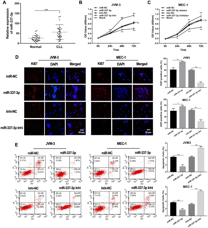
Figure 1.
Expression and functions of miR-337-3p in CLL. (A) Relative expression levels of miR-337-3p in PBMC samples from thirty treatment-naïve CLL patients and thirty normal donors. (B, C) CCK8 assay was performed to assess the influence of miR-337-3p on CLL cells proliferation. (D) IF assay analyzed the proliferative viability of JVM-3 and MEC-1 after transfected with miR-337-3p mimics and inhibitor and ki67-positive cells rate were calculated. (E) Cell apoptosis was detected by FCM to verify the effects of miR-337-3p and the percentage of apoptotic cells was quantified. Three biological replicates were performed per condition and mean values ± SD are displayed (*P < 0.05; **P < 0.01; ***P < 0.001; ns, not significant).

