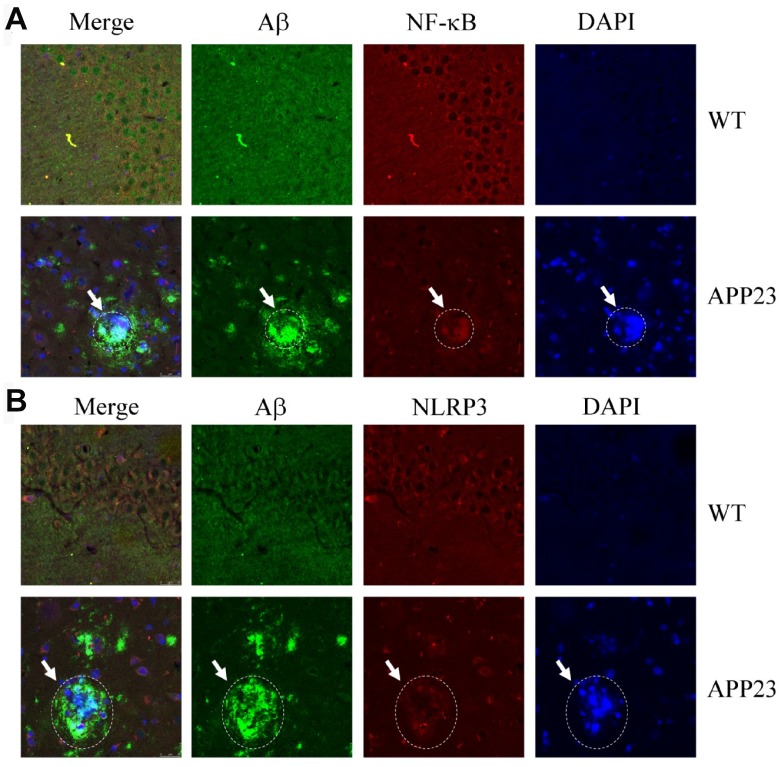
Figure 2.
Confocal fluorescence microscopy shows colocalization of NF-κB, NLRP3, and Aβ42 in the brains of APP23 mice. Images from a single z-plane of the brains of APP23 mice at 6 months of age using monoclonal antibodies against Aβ (green), NF-κB/NLRP3 (red), and nucleus (blue DAPI stain). (A) Colocalization of NF-κB and Aβ42 is observed as indicated in the circle. (B) Colocalization of NLRP3 and Aβ42 was observed as indicated in the circle. Scale bar=25 μm.

