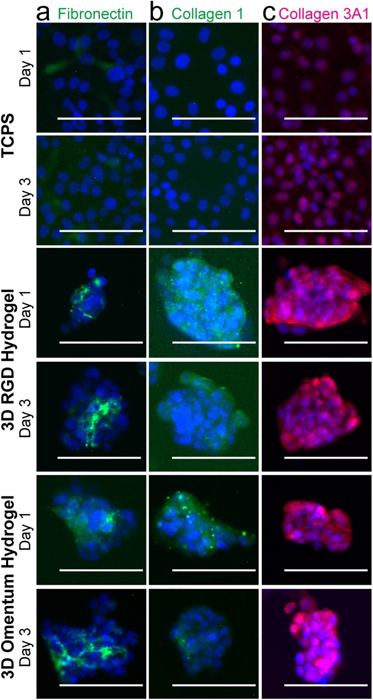
FIG. 4.
Immunofluorescence staining of SKOV-3 cells demonstrated that they produce their own ECM proteins over the time frame of a drug screening assay. (a) Fibronectin (green)/DAPI (blue). Fibronectin can be seen on plastic as well as between cells in the MCTS. (b) Collagen 1 (green)/DAPI (blue). Collagen 1 production was not observed on plastic but was visible around the spheroids. (c) Collagen3A1 (pink)/DAPI (blue). Some collagen 3A1 may be present on plastic but is clearly visible surrounding the spheroids in both hydrogels. Scale bars = 100 μm. Images are representative from N ≥ 2 independent experiments.

