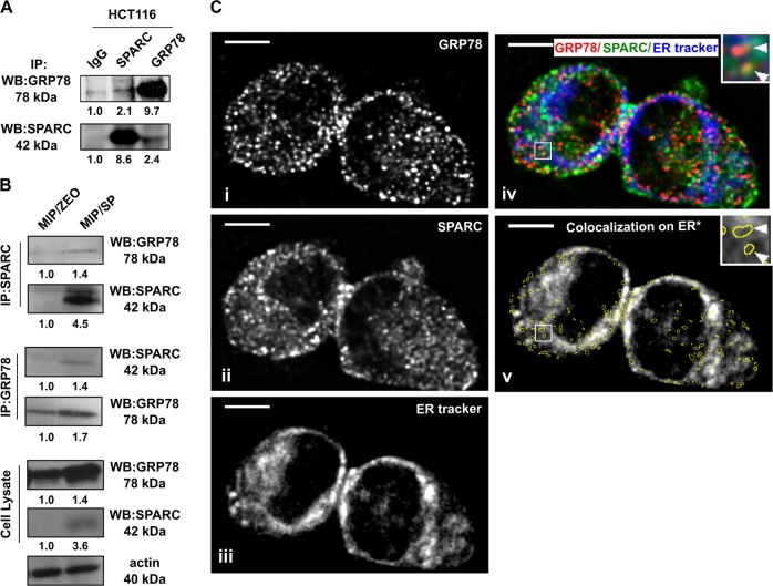
Fig. 1. Interaction between GRP78 and SPARC in CRC.
Co-immunoprecipitation of SPARC with GRP78 in (a) HCT116 and confirmed in (b) MIP/Zeo and MIP/SP cells. (c) Colocalization of (i) GRP78 and (ii) SPARC in HCT116 by confocal microscopy and immunofluorescent analysis. (iii) The endoplasmic reticulum was stained with ER tracker blue-white DPX dye. (iv) Images were overlapped, and (v) the GRP78:SPARC colocalization pixels were identified by Colocalization Finder plugin in ImageJ and highlighted with yellow circles. The average ER tracker staining intensity in the highlighted areas was calculated to determine the percentage of GRP78:SPARC colocalization in the ER. Scale bar = 5 μm

