
Figure 1.
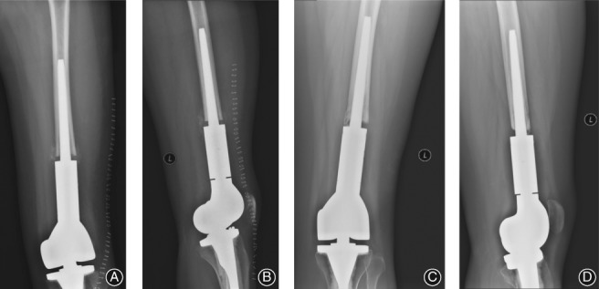
Biologic fixation of a distal femoral prosthesis in a 24‐year‐old woman following resection of an osteosarcoma. Frontal (A) and lateral (B) radiographs showing an adequate contact between the prosthetic stem and the medial side of the cortical bone in the medullary cavity, with a visible 1–2 mm space between the prosthesis stem and the bone. Repeat frontal (C) and lateral (D) radiographs obtained 54 months post‐implantation, showing good contact between the prosthesis and the medullary cavity and satisfactory fixation, with the space between the bone and the prosthesis being maintained, with formation of a bony bridge.