
Figure 5.
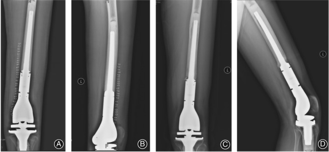
Aseptic loosening of the distal femoral component in a 14‐year‐old male treated for an osteosarcoma using cemented fixation of the distal prosthesis. Frontal (A) and lateral (B) views of the distal femoral component after surgery show a centralized positioning of the stem, with a translucent band visible between the cement layer of the proximal prosthesis stem and the cortical bone. Frontal (C) and lateral (D) view of the distal femur obtained 37 months after the surgery, with the translucent band being larger than on the initial baseline radiographs obtained post‐surgery.