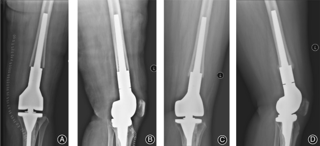
Figure 6.

Aseptic loosening in a 40‐year‐old woman treated for a chondrosarcoma, using biologic fixation of the prosthesis. Frontal (A) and lateral (B) views of the distal femur, obtained after surgery, with a short contact length between the stem and the cortical bone of the medullary cavity, with absence of a 3‐point fixation and no visible space between the stem and the medullary cavity. Frontal (C) and lateral (D) view of the distal femur obtained 8 months after prosthesis implantation. The prosthesis stem could not form an effective fixation with the medullary cavity, with upward migration of the prosthesis and the patient complaining of pain.
