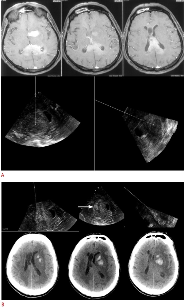
Fig. 1. Navigated 3D ultrasound-guided biopsy of a 70-year-old man with multiple intracranial lesions.

A. The upper panel shows axial T1-weighted magnetic resonance imaging with multiple homogenously enhancing lesions in the left gangliocapsular region, body, and splenium of the corpus callosum. The lower panel shows real-time intraoperative images obtained using 3-dimensional navigated ultrasound (two planes depicted side by side). The solid line represents the navigator, and the dotted lines represent the virtual offset planned for targeting the lesion for biopsy, along which the same navigator-mounted needle is passed for biopsy. B. Intraoperative navigated ultrasound images (in three orthogonal planes) taken after biopsy of the lesion, show a small hematoma (arrow) in the tumour bed, along with track hematoma. Postoperative computed tomography images show a hematoma at the biopsy site (corresponding to the ultrasound images), which was managed conservatively.
