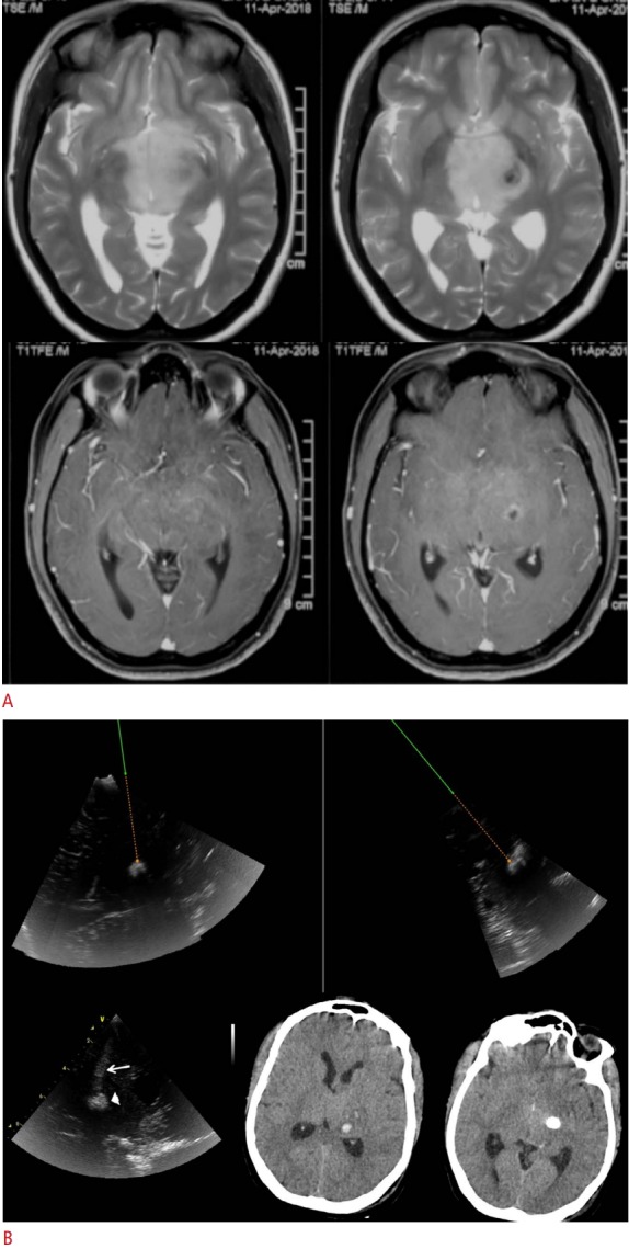
Fig. 2. Navigated 3D ultrasound-guided of a 35-year-old woman with bithalamic masses.

A. Axial T2-weighted (top row) and postgadolinium contrast (bottom row) magnetic resonance images show diffuse and faintly enhancing lesions involving the bilateral thalami, with foci of suspected necrosis/ calcification in the left gangliocapsular lesion. B. Intraoperative ultrasound images of the same case are shown. The top row shows intraoperative ultrasound (navigated ultrasound) images in two planes, in which the target is set with an offset, and the virtual track along which the navigator-mounted needle is passed for biopsy. The lower post-biopsy ultrasound image (left) shows a small hematoma in the biopsy bed (arrowhead) and along the biopsy track (arrow) seen immediately after targeting the biopsy. Postoperative computed tomography scans show the small hematoma at the biopsy site (lower middle) and the deeper residual calcification (lower right). The hematoma was asymptomatic and conservatively managed.
