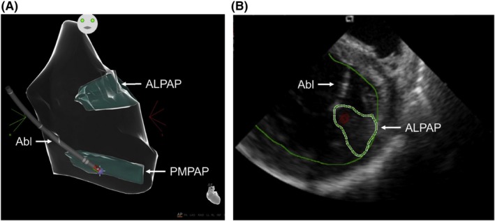
Figure 8.

Intraprocedural imaging during ablation of papillary muscle arrhythmias. A, Anatomical map of the left ventricle (CARTO, Biosense Webster) showing contact of the ablation catheter (Abl) with the posteromedial papillary muscle (PMPAP). B, Intracardiac echocardiogram showing real‐time visualization of the ablation catheter during ablation on the anterolateral papillary muscle (ALPAP)
