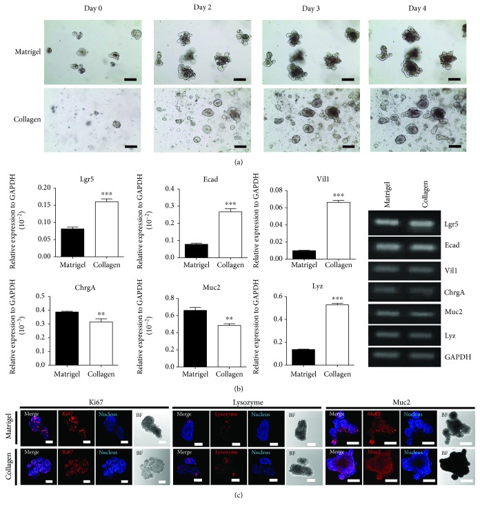
Figure 3.
Comparison of growing mouse small intestinal organoids in Matrigel and in collagen-based matrix culture system. (a) Growth pattern and morphology of mouse small intestinal organoids in both Matrigel and collagen-based matrix during Passage 1. Bar scale is 50 μm. (b) Quantitative RT-PCR analysis of the markers of intestinal organoids such as Lgr5, Ecad, Vil1, ChrgA, Muc2, and Lyz at Passage 4 cultured in both matrices (left). Gene expression shown as gel electroporation (right). ∗∗p < 0.01, ∗∗∗p < 0.001. (c) The immunocytochemistry staining of Ki67, mucin 2, and lysozyme was examined in Matrigel and Collagen matrix. Scale bar: 100 μm.

