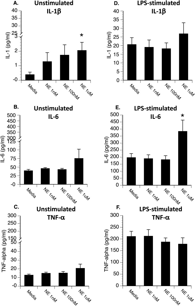
Figure 3.
Hippocampal microglia were plated in a 96-well culture plate at 10,000 cells per well and incubated with either (A–C) media (unstimulated) or (D–F) 0.5 μg LPS (stimulated), along with different concentrations of norepinephrine. After 20 hours, supernatants were collected for measurement of IL-1β, IL-6, and TNF-α by ELISA (R&D Systems). Under unstimulated conditions (A–C), norepinephrine resulted in (A) a dose-dependent increase in IL-1β (A). Under LPS-stimulated conditions (D–F), the highest concentration of norepinephrine significantly increased (E) LPS-induced IL-6 release. *P < 0.05 difference from media-treated cells.

