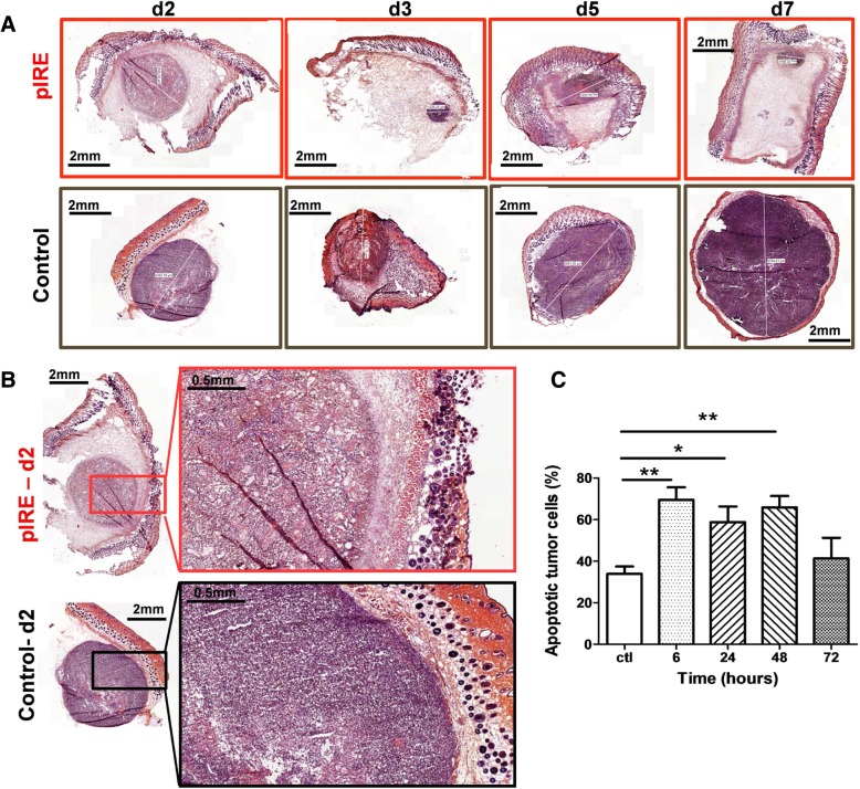
Fig. 2.
pIRE induced tumor apoptotic cell death. C57Bl/6 mice were intradermally injected with 0.5 × 10⁶ B16F10 cells. When the tumor reached a volume of 20 to 30 mm3, pIRE parameters were applied: 10 square waved pulses of 1200 V, duration 100 μs, frequency 1 kHz. A and B-Tumor were collected from 2 to 7 days after pIRE treatment. After fixation and inclusion, 5 μm sections were cut, stained with eosin and hematoxylin, mounted and observed with a scanner. a Eosin-hematoxylin representative images of skin tumors 2 (d2), 3 (d3), 5 (d5) and 7 days (d7) after pIRE treatment (up line) or without treatment (bottom line). Tumor diameter is highlighted by a white line. b High magnification, 2 days after pIRE, of representative images of skin tumors treated (top) or not (bottom). c Tumors were collected 6, 24, 48 and 72 h after pIRE treatment, dissociated, and cells suspensions labelled with Annexin V-FITC and 7AAD to characterize cell death. Histograms of Annexin V positive/7AAD negative cells described as early apoptotic in untreated mice (CTL) and 6 to 72 h post pIRE treatment. For untreated mice, a tumor from each time point was collected. Values are means ± SEM of 4 mice. * < 0.05 and **P < 0.01 (Two-way ANOVA analysis)

