Fig. 3.
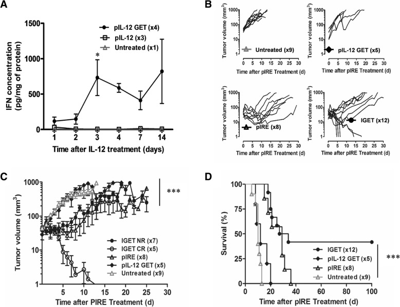
IGET treatment induced partial B16F10 tumor regression. a CD11c-GFP-DTR mice were intradermally injected with 25 μg of pIL-12 in 20 μl PBS, or with 20 μl of PBS alone in one flank and were submitted to GET protocol. The contralateral flank from IL-12 injected mice was non injected and non-pulsed to be used as an internal control. Mice were sacrificed from 1 to 14 days after treatment and skin from each flank were harvested and cultured overnight in complete medium 5%CO2, 37 °C. IFN-γ content was determined in the cultured supernatant of pIL-12 treated skin (pIL-12 GET), of pIL-12 untreated skin (pIL-12) and of PBS treated skin (PBS GET) by ELISA. 3 ≤ n ≤ 4, 2 independent experiments, statistical analysis: 2way Anova, *p < 0.05. b CD11c-GFP-DTR mice were intradermally injected with 0.5 × 10⁶ B16F10 cells. When the tumor reached a volume of 7 to 15 mm3, 2x25μg of pIL-12 were injected intradermally in the healthy skin directly surrounded the tumor. Electrotransfection was realized using GET protocol. Two days later, pIRE parameters were applied on previously treated tumors. Tumor volume was followed up to 100 days in case of complete regression. Individual curves are plotted for untreated control, pIL-12-GET alone, pIRE alone, and IGET treated mice. c Mean +/− SEM tumor growth progression while separating IGET treated group between complete regression group (CR) and non-responding group (NR). From day 4, ***p < 0.001 between IGET CR and untreated or pIL-12-GET groups (Two-way ANOVA analysis). d Mice survival of untreated control, pIL-12 GET, pIRE and IGET treated tumor. ***P < 0.001 (Log-rank (Mantel-Cox) Test) between untreated control, pIL-12 GET, pIRE and IGET treated tumor (5 ≤ n ≤ 12, 3 independent experiments)

