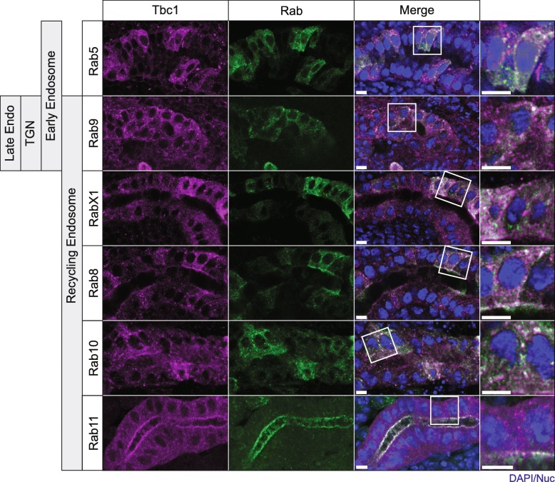
Fig. 4.
Partial Colocalization of Tbc1 with a subset of Rabs. Tbc1: purple; Rab: Green; DAPI: Blue. UAS-tbc1 and UAS-YFP-Rab were co-expressed in the SG using a fkh-Gal4 driver on chromosome II that has mosaic expression except for the Rab11 stain (bottom). Anti-Tbc1 was used to detect Tbc1 and anti-GFP was used to detect the Rab proteins in the top five sets of experiments. For Rab11 costaining, endogenous Rab11 was detected with Rab11 antiserum in a fkh-Gal4(III) > UAS-tbc1-GFP background. In this instance, GFP antibody was used to detect Tbc1. Rabs are organized according to Flybase annotations [44]. Scale bar: 5 μm

