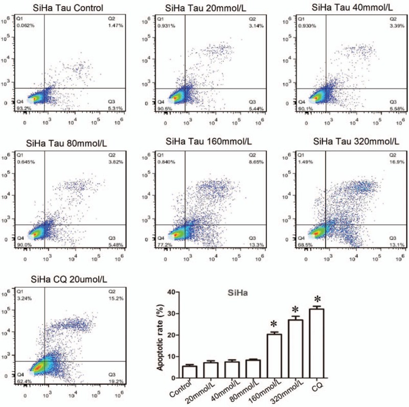
Figure 2.

Effects of Tau on the apoptosis of cervical cancer cells. The SiHa cells were treated by Tau with different concentrations 0 (control), 20, 40, 80, 160, 320 mmol/L Tau, and 20 μmmol/L DDP for 48 h, and the apoptosis of SiHa cells was detected by the Annexin-V-FITC/PI double staining method. DDP is the positive control. ∗P < 0.01 vs. the Control group.
