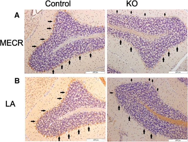
Figure 1.
MECR and LA abundance in cerebellar sections of control and KO mice at 6 months of age. The sections were treated with antibodies either to MECR or lipoylated proteins and counterstained with hematoxylin. A, Compared with the control samples (left) Mecr PCs of 6-month-old Pcp2-Cre KO mice (right) appeared to elicit lower intensities for the MECR antibody signal, with generally weaker brown staining, indicating reduced MECR protein levels. B, Anti-LA staining of cerebellar sections. Compared with control samples (left), the tissue sections of KO mice cerebella (right) treated with anti-LA antibody display a mosaic staining pattern congruent with the MECR pattern in the panel above, indicating that levels of lipoylated proteins were also reduced in PCs of KO mice. Arrows point to examples of PCs. Scale bar, 200 μm.

